Risk factors for the development of congenital heart defects in Kyrgyz Republic
ORIGINAL RESEARCH ARTICLE
Risk factors for the development of congenital heart defects in Kyrgyz Republic
Article Summary
- DOI: 10.24969/hvt.2026.629
- CARDIOVASCULAR DISEASES
- Published: 03/03/2026
- Received: 30/01/2026
- Revised: 01/03/2026
- Accepted: 01/03/2026
- Views: 1143
- Downloads: 976
- Keywords: Congenital heart defects, risk factors, demographic factors, biological factors, chemical factors pediatric cardiology
Address for Correspondence: Sоfia M. Shakhnabieva, Medical Faculty, Kyrgyz-Russian Slavic University, Bishkek, Kyrgyzstan
E-mail: shahnabieva_sofia@mail.ru
ORCID: Sоfia M. Shakhnabieva - 0000-0002-6811-9580; Shakhsianam Sh. Khasanova- 0009-0008-0101-7505;
Elena I. Kondrateva - 0000-0002-0674-4903.
Sоfia M. Shakhnabieva, Shakhsianam Sh. Khasanova, Tatiana I. Sologubovа, Elena I. Kondrateva, Ainagul D. Niiazalieva, Yuri M. Kurmanbakeev
Medical Faculty, Kyrgyz-Russian Slavic University, Bishkek, Kyrgyzstan
Abstract
Objective: Congenital heart diseases (CHD) are structural abnormalities of the heart and/or great vessels that are present at birth. CHD is still the most common inborn defect with an approximate prevalence of 5–11 per 1000 live births and incidence of 1%. CHDs are caused by many factors, often a mix of genetic predispositions and environmental influences, though the specific cause is unknown for most cases.
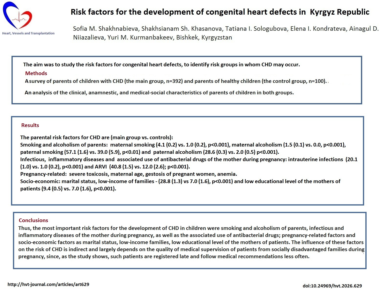
The aim was to study the risk factors for congenital heart defects, to identify risk groups in whom CHD may occur.
Methods: We conducted a survey of parents of children with CHD (the main group, n=392) and parents of healthy children (the control group, n=100), followed by an analysis of the clinical, anamnestic, and medical-social characteristics of parents of children in both groups. The following groups of risk factors were identified: demographic factors, socio-hygienic factors, biological factors, and chemical factors.
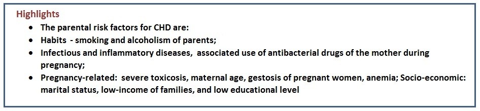
Results: The most important risk factors for the development of CHD in children were bad habits of parents: maternal smoking and alcoholism ((4.1 (0.2) vs. 1.0 (0.2), p<0.001, main group vs control; (1.5 (0.1) vs. 0.0, p<0.001, respectively), paternal smoking (57.1 (1.6) vs. 39.0 (5.9), p<0.01), paternal alcoholism (28.6 (0.3) vs. 2.0 (0.5) p<0.001); infectious and inflammatory diseases of the mother during pregnancy: acute respiratory viral infections (40.8 (1.5) vs. 12.0 (2.6); p<0.001, main vs. control group), intrauterine infections (20.1 (1.0) vs. 1.0 (0.2), p<0.001); as well as the associated use of antibacterial drugs. Conditions directly related to pregnancy (severe toxicosis, maternal age, gestosis of pregnant women, anemia) made a less noticeable but significant contribution to determining the risk. The increased risk of having a child with CHD was determined by a number of socio-economic factors: marital status, low-income families (28.8 (1.3) vs 7.0 (1.6) main vs control group, p<0.001) and low educational level of the mothers of patients (9.4 (0.5) vs. 7.0 (1.6), p<0.001).
Conclusion: Thus, the most important risk factors for the development of CHD in children were smoking and alcoholism of parents, infectious and inflammatory diseases of the mother during pregnancy, as well as the associated use of antibacterial drugs; pregnancy-related factors as severe toxicosis, maternal age, gestosis of pregnant women, anemia; and socio-economic factors as marital status, low-income families, low educational level of the mothers of patients. The influence of these factors on the risk of CHD is indirect and largely depends on the quality of medical supervision of patients from socially disadvantaged families during pregnancy, since, as the study shows, such patients are registered late and follow medical recommendations less often.
Key words: Congenital heart defects, risk factors, demographic factors, biological factors, chemical factors pediatric cardiology
Introduction
Congenital heart diseases (CHDs) are structural abnormalities of the heart and/or great vessels that are present at birth (1- 3). CHDs include an extensive range of defects that may be minor and require no intervention or may be life-limiting and require complex surgery shortly after birth (4, 5). CHD is still the most common inborn defect with an approximate prevalence of 5–11 per 1000 live births and incidence of 1% (6).
CHD happen because the heart does not develop normally while the baby is growing in the womb.
Graphical abstract
Genetics plays an important role in the formation of CHD (7, 8). It is common for CHD to occur because of changes, or mutations in the child’s deoxyribonucleic acid (DNA) (2). The mutations in the DNA may or may not have come from the parents (9, 10).
Rarely, CHD are caused by particular genes that are Inherited from the parents. That means a parent who has a CHD may be more likely to have a child with the defect (11-13). It is also known that fetuses with a prenatal diagnosis of CHD have an increased risk of several comorbidities, including intrauterine growth restriction (14).
Previously, similar studies were conducted in Kyrgyzstan, but they did not include various groups of risk factors, and they were not comprehensive, so to undertake study on risk factors of CHD is timely and relevant.
The aim was to study the risk factors for congenital heart defects, to identify risk groups in whom CHD may occur.
Methods
Study design and population
This is a cross-sectional survey study. To study the main risk factors for having a child with CHD, parents of children with CHD (the main group, n=392) and parents of healthy children (the control group, n=100) were surveyed, followed by an analysis of the clinical, anamnestic, and medical-social characteristics of parents of children in both groups.
The survey was conducted in 19 family medicine centers from January 2024 to January 2025 in Bishkek, Kyrgyzstan among the main group of 392 patients with CHD: 120 patients with CHD (from 18 years old), 272 parents of children with CHD (under 18 years old), according to questionnaires developed by us in Russian and Kyrgyz languages. Inclusion criteria were: age 1 month - 60 years, diagnosed with «Congenital heart disease» (anatomical abnormalities of the heart/blood vessels), signed informed consent. Exclusion criteria: 1) severe concomitant pathology: diseases that are a contraindication to cardiac surgery; 2) absence of indications: defects that do not require active treatment or correction; 3) chromosomal/extracardial disorders: severe genetic syndromes that limit life if it is not a subject of study; 4) other reasons: infectious diseases or refusal to participate.
The data of 100 parents of healthy children without heart murmur who were evaluated in the children's department of maternity hospital No. 1 in Bishkek were used as a comparison group. The condition at birth of all children was assessed on the Apgar scale of at least 9 points, without signs of disability, having the first group of health.
Informed consent to participate in survey was signed by the parents/legal representatives and the patient (if >18 years old). The study was conducted according to the rules of the Helsinki 2024 agreement and approval of Institutional Ethics Committee was obtained.
Risk factors of CHD
We collected the following data:
• Demographic factors (gender, age, marital status, education level);
• Social and hygienic factors (material and living conditions in the family, bad habits (smoking, alcoholism, drug addiction, working at a personal computer, using a smartphone);
-Diseases in close relatives (diseases of the cardiovascular system, a history of congenital anomalies, CHD in the immediate family);
-Obstetric history (number of pregnancies, abortions, childbirths, features of the course of a real pregnancy (toxicosis, gestosis, bleeding, fetal infections));
- Diseases, mothers suffered during pregnancy (acute respiratory viral infection (ARVI), anemia, pyelonephritis, vaginitis, urethritis, colpitis, hypertension, diabetes, infection);
• Chemical factors (taking medications during pregnancy).
Survey
The survey was conducted through specially designed questionnaires in Russian and Kyrgyz languages. The questionnaires were distributed among the 19 Bishkek city family medicine centers, and they were handed over to pediatricians and internal medicine physicians, where patients live at the district and are monitored by these doctors. Parents of patients with CHD answered the questions. Then we collected the questionnaires and analyzed them. We did not interview patients from the country's regions.
During the survey, World Health Organization (WHO) data were used to determine smoking: about 200 puffs of smoke from smoldering tobacco leaves per day, which is 1 pack of cigarettes per day (15). Alcoholism was defined as a clinically established diagnosis, according to WHO: "a primary, chronic disease characterized by impaired control over alcohol intake, addiction to alcohol, alcohol consumption, despite the negative consequences." (16).
To determine family income, the average subsistence minimum in the Kyrgyz Republic was used (17), amounting to 8 758,6 soms (for 2025), low–income families had incomes in the family below the average subsistence minimum, middle-income families had incomes at the level of the average subsistence minimum, and families with high incomes were defined as having incomes above the average subsistence minimum.
When we studied the use of various medications, it turned out that medications were taken at different stages of pregnancy. Most of the women took various preventive medications before the 13th week of pregnancy (Iodomarin, folic acid, multivitamins).
Those who had iron deficiency anemia took iron supplements throughout pregnancy. The women took actovegin, calcium gluconate, antibiotics, nonsteroidal anti-inflammatory agents, and other medications in courses, depending on when the need arose.
Statistical analysis
Statistical data processing was performed using Microsoft Excel spreadsheets and the Statistica for Windows v. 10.0 application software package from StatSoft Inc. (USA), as well as the Biostat statistical program. Statistical processing of the materials was carried out using modern parametric and nonparametric methods of statistical analysis of biomedical research, as well as criteria of evidence-based medicine in accordance with modern requirements for the analysis of medical data. The representativeness was determined according to the generally accepted methodology of A.M. Merkov (1974) with an unknown number of the general population.
The statistical method of the study included the calculation of intensive, extensive indicators, the error of representativeness (± m), the criterion of reliability (t), the effectiveness of error-free prediction (p=95.0%; p=99.9%, with p<0.01; p<0.001), dynamic series (absolute increase, growth rate), correlation.
Results
The level of wealth and marital status in the families of children with CHD in the study groups is presented in Table 1.
|
Table 1. The level of wealth and marital status in families of children with CHD (per 100 respondents) in the study groups |
||||||
|
# |
Level of wealth and marital status of parents |
Children with CHD Main group (n= 392) |
Healthy children Control group (n= 100) |
t |
||
|
the abs. number |
Р (m) |
the abs. number |
Р (m) |
p |
||
|
1. |
High income |
36 |
9.2 (0.5) |
12 |
12.0 (2.7) |
1.5 |
|
>0.05 |
||||||
|
2. |
Medium income |
243 |
62.0 (1.5) |
81 |
81.0 (1.0) |
23.8 |
|
<0.001 |
||||||
|
3. |
Low income |
113 |
28.8 (1.3) |
7 |
7.0 (1.6) |
20.5 |
|
<0.001 |
||||||
|
4. |
Married |
221 |
56.4 (1.6) |
87 |
87.0 (2.8) |
11.8 |
|
<0.001 |
||||||
|
5. |
Not married |
156 |
39.8 (1.5) |
13 |
13.0 (2.8) |
10.7 |
|
<0.001 |
||||||
|
6. |
No answer |
15 |
3.8 (0.2) |
- |
- |
- |
|
P (m) is an intensive indicator and an error of representativeness; t is reliability; p is the effectiveness of an error–free forecast (with p=95.0%, 99.9%) abs. – absolute, CHD - congenital heart disease |
||||||
As follows from the data presented in Table 1, children in families with unfavorable socio-hygienic factors, such as family poverty (p<0.001), parents are not married (p<0.001), poor living conditions in the family were significantly more likely to suffer from CHD. At the same time, there was no increase in the incidence of CHD in families with good conditions (p>0.05).
The level of education and employment of parents in children with congenital heart disease is presented in Table 2.
When studying the influence of the level of education and employment of parents on the risk of developing CHD in children, the following results were obtained. It turned out that the father's higher education (p<0.001) had a protective effect and reduced the risk of developing CHD.
A low level of education in the mother increased the risk of developing CHD in the child (p<0.001). The employment of the father reduced the risk of developing CHD in the child (p<0.001). The employment of the mother did not have a significant impact on the risk of developing CHD in children (p>0.05) (Table 2).
|
Table 2. The level of education and employment of parents in children with CHD |
||||||
|
Variables |
Categories of education and employment |
Children with CHD Main group (n= 392) |
Healthy children Control group (n= 100) |
t/p |
||
|
the abs. number |
Р (m) |
the abs. number |
Р (m) |
|||
|
Father's education |
Average |
203 |
51.8(1.6) |
33 |
33.0(5.5) |
2.2(<0.01) |
|
Higher |
126 |
32.1(1.4) |
67 |
67.0(5.5) |
4.3(<0.001) |
|
|
Primary |
57 |
14.5(0.8) |
0 |
- |
- |
|
|
Mother's education |
Average |
215 |
54.8(1.6) |
58 |
58.0(6.0) |
* >0.05 |
|
Higher |
140 |
35.7(1.5) |
35 |
35.0 (5.7) |
* >0.05 |
|
|
Primary |
37 |
9.4(0.5) |
7 |
7.0(1.6) |
3.4(<0.001) |
|
|
Father's employment |
Yes |
298 |
76.0(1.1) |
86 |
86.0(3.0) |
4.0(<0.001) |
|
No |
82 |
20.9(1.0) |
14 |
14.0(3.0) |
2.3(<0.01) |
|
|
Mother's employment |
Yes |
137 |
34.9(1.4) |
39 |
39.0(5.9) |
* >0.05 |
|
No |
255 |
65.0(1.5) |
61 |
61.0(5.9) |
* >0.05 |
|
|
P (m) is an intensive indicator and an error of representativeness; t is reliability; p is the effectiveness of an error–free forecast (with p=95.0%, 99.9%) abs. – absolute, CHD - congenital heart disease |
||||||
Data on mother`s age and cardiovascular disease in relatives of patients with CHD are presented in Table 3.
|
Table 3.Mother's age during pregnancy, cardiovascular diseases in close relatives of patients with CHD |
|||||||
|
# |
Variables |
Children with CHD main group (n= 392) |
Healthy children control group (n= 100) |
Reliability |
Interconnection (correlation ) |
||
|
abs. number |
Р (m) |
abs. number |
Р (m) |
t/p |
r xy |
||
|
Mother's age, years |
|||||||
|
1. |
From 16 to 20 |
45 |
11.5 (0.6) |
13 |
13.0(2.8) |
*/ >0.05 |
The connection is direct, strong +1,0 |
|
2. |
21-30 |
182 |
46.4(1.6) |
61 |
61.0(5.9) |
*/ >0.05 |
|
|
3. |
From 31 to 45 |
165 |
42.1(1.5) |
26 |
26.0(4.8) |
6.3 /<0.001 |
|
|
Cardiovascular diseases in close relatives of patients with CHD |
|||||||
|
4. |
No |
272 |
69.4(1.3) |
78 |
78.0(4.3) |
*/>0.05 |
No connection |
|
5. |
Yes |
120 |
30.6(1.3) |
22 |
22.0(4.3) |
*/ >0.05 |
|
|
P (m) is an intensive indicator and an error of representativeness; t is reliability; p is the effectiveness of an error–free forecast (with p=95.0%, 99.9%)* – there is no significant difference, r xy is a correlation relationship. abs. – absolute, CHD - congenital heart disease |
|||||||
From the data presented in Table 3, it follows that the age of the mother has a significant impact on the risk of CHD in children. In particular, the age of the mother over 30 years increases the likelihood of developing CHD in a child (p<0.001). The correlation is direct and strong, +1.0. At the same time, there was no increase in the risk of having children with CHD in mothers less than 20 years of age (Table 3). It should also be noted that burdened inheritance of cardiovascular diseases from both the mother and father does not significantly increase the risk of having a child with CHD (p>0.05).
|
Table 4. Prevalence and influence of risk factors in parents on the development of CHD |
||||||||||
|
# |
Risk factors |
Parents with risk factors (n= 784) |
p |
Interconnection (correlation )r xy |
||||||
|
Main group |
Control group |
|||||||||
|
abs. number |
Р (m) |
abs. number |
Р (m) |
|||||||
|
1. |
Mother's smoking |
16 |
4.1(0.2) |
1 |
1.0(0.2) |
<0.001 |
The connection is direct, strong (+1,0)
|
|||
|
2. |
Father's smoking |
224 |
57.1(1.6) |
39 |
39.0(5.9) |
<0.01 |
||||
|
3. |
Mother's alcoholism |
6 |
1.5 (0.1) |
0 |
0.0 |
<0.001 |
||||
|
4. |
Father's alcoholism |
112 |
28.6(0.3) |
2 |
2.0(0.5) |
<0.001 |
||||
|
5. |
A mother's job on a PC |
123 |
31.4(1.3) |
11 |
11.0(2.4) |
* >0.05 |
||||
|
6. |
Phone usage (a cell phone (or mobile phone) |
280 |
71.4 (1.3) |
71 |
71.0(5.1) |
* >0.05 |
||||
|
P (m) is an intensive indicator and an error of representativeness; t is reliability; p is the effectiveness of an error–free forecast (with p=95.0%, 99.9%)* – there is no significant difference, r xy is a correlation relationship abs. – absolute, CHD - congenital heart disease, PC – personal computer |
||||||||||
When assessing the effect of bad habits in parents on the risk of developing CHD in a child (Table 4), it turned out that maternal smoking (p<0.001), maternal alcoholism (p<0.001), paternal smoking (p<0.01), paternal alcoholism (p<0.001) significantly increases the risk of CHD in children. The correlation is direct, strong, +1.0. At the same time, the mother's work on a personal computer (PC) and the use of a cell phone did not significantly affect the risk of developing CHD (p>0.05) (Table 4).
|
Table 5. Obstetric history and features of pregnancy in women of the main and control groups (per 100 respondents) |
|||||||
|
N |
Obstetric history |
Mothers with a risk factor (n= 392) |
p |
Interconnection (correlation )rxy |
|||
|
Main group |
Control group |
||||||
|
abs. number |
Р (m) |
abs. number |
Р (m) |
||||
|
1. |
Abortions |
112 |
28.6(1.3) |
27 |
27.0(4.9) |
* >0.05 |
The connection is direct, strong (+1,0) |
|
2. |
Severe toxicosis |
275 |
70.2(1.4) |
48 |
48.0(6.2) |
<0.001 |
|
|
3. |
ARVI |
160 |
40.8(1.5) |
12 |
12.0(2.6) |
<0.001 |
|
|
4. |
Gestosis |
148 |
37.8(1.5) |
13 |
13.0(2.3) |
<0.001 |
|
|
5. |
IUI |
79 |
20.1(1.0) |
1 |
1.0(0.2) |
<0.001 |
|
|
6. |
Bleeding |
27 |
6.9(0.4) |
6 |
6.0(1.4) |
* >0.05 |
|
|
7. |
Anemia |
49 |
12.5(0.7) |
3 |
3.0(0.7) |
<0.001 |
|
|
8. |
Pyelonephritis |
47 |
12.0(0.7) |
6 |
6.0(1.4) |
<0.001 |
|
|
9. |
Vaginitis |
3 |
0.8(0.05) |
1 |
1.0(0.2) |
<0.01 |
|
|
10 |
Urethritis |
4 |
1.0(0.06) |
1 |
1.0(0.2) |
* >0.05 |
|
|
11. |
Colpitis |
33 |
8.4(0.5) |
4 |
4.0(1.0) |
<0.001 |
|
|
P (m) is an intensive indicator and an error of representativeness; t is reliability; p is the effectiveness of an error–free forecast (with p=95.0%, 99.9%)* – there is no significant difference, r xy is a correlation relationship abs. – absolute, ARVI - acute respiratory viral infections, IUI - Intrauterine infection |
|||||||
An analysis of obstetric history data showed that the presence of previous abortions and the number of pregnancies had no significant effect on the risk of CHD in children (p>0.05) (Table 5). The most pronounced effect on the risk of developing CHD in a child was exerted by pathological conditions that develop during pregnancy. Thus, the presence of intrauterine infection (IUI) significantly increased the risk of CHD in children (, p<0.001), as well ARVI disease (p<0.001), anemia (p<0.001), gestosis (p<0.001), and severe toxicosis (p<0.001). The correlation is direct and strong (+1.0). At the same time, we did not find an association between the presence of inflammatory diseases of the urinary tract and bleeding in the mother with an increased risk of CHD in children (p>0.05) (Table 5).
|
Table 6. Frequency of taking medications during pregnancy (per 100 respondents) |
||||||
|
N |
Drugs
|
Frequency of taking the drug |
t/p
|
|||
|
Main group |
Control group |
|||||
|
abs. number |
Р (m) |
abs. number |
Р (m) |
|||
|
1. |
Iron-containing preparations |
35 |
8.9(0.5 |
13 |
13.0(2.8) |
2.0 /<0.01 |
|
2. |
Iodomarin |
12 |
3.1(0.2) |
3 |
3.0(0.7) |
*/>0.05 |
|
3. |
Multivitamins |
23 |
5.9(0.3) |
4 |
4.0(1.0) |
7.0/<0.001 |
|
4. |
Actovegin |
3 |
0.8(0.05) |
2 |
2.0(0.5) |
20.0/ <0.001 |
|
5. |
Calcium gluconate |
8 |
2.0(0.1) |
1 |
1.0(0.2) |
10.0/ <0.001 |
|
6. |
Folic acid |
9 |
2.3(0.1) |
3 |
3.0(0.7) |
10.0/ <0.001 |
|
P (m) is an intensive indicator and an error of representativeness; t is reliability; p is the effectiveness of an error–free forecast (with p=95.0%, 99.9%)* – there is no significant difference, r xy is a correlation relationship |
||||||
A study was conducted on the effect of pregnant women taking a number of medications (iron-containing, iodomarin, multivitamins, folic acid, calcium gluconate, and actovegin) on the risk of CHD in children. It turned out that taking multivitamins (p<0.001) and calcium gluconate (p<0.001) increases the risk of CHD. Taking iron-containing drugs, folic acid, and actovegin was not associated with an increased risk of having children with developmental abnormalities of the cardiovascular system. However, these data should be interpreted with caution due to the small number of such observations in both the main and control groups (Table 6).
At the same time, a relationship has been established between the birth of children with CHD and taking medications for infections during pregnancy (Table 7).
|
Table 7. Frequency of medications taken for acute respiratory viral infections during pregnancy (per 100 respondents) |
||||||
|
N
|
Medications |
Frequency of taking the drug |
||||
|
Main group |
Control group |
t/p
|
||||
|
abs. number |
Р (m) |
abs. number |
Р (m) |
|||
|
1. |
Antipyretics |
30 |
7.7(0.4) |
8 |
8.0(1.8) |
*/>0.05 |
|
2. |
Expectorants |
19 |
4.8(0.3) |
2 |
2.0(0.5) |
10.0/ <0.001 |
|
3. |
Antihistamines |
8 |
2.0(0.1) |
- |
- |
- |
|
4. |
Antiviral drugs |
13 |
3.3(0.2) |
- |
- |
- |
|
5. |
NSAIDs |
23 |
5.9(0.3) |
1 |
1.0(0.2) |
16.3/<0.001 |
|
6. |
Antifungal |
14 |
3.6(0.2) |
- |
- |
- |
|
7. |
Antibacterial |
107 |
27.3(1.3) |
4 |
4.0(1.0) |
25.8/<0.001 |
|
P (m) is the intensive indicator and the error of representativeness; T is the reliability; P is the effectiveness of an error–free forecast (at p=95,0%, 99,9%), * – there is no significant difference NSAIDs – nonsteroidal anti-inflammatory drugs |
||||||
Thus, the use of antibacterial agents by women for intrauterine infections (p<0.001), nonsteroidal anti-inflammatory drugs (p<0.001), expectorants was associated with an increased risk of childbirth with the CHD. Antibacterial drugs taken by pregnant women were of various groups, such as penicillins (ampicillin, amoxicillin, amoxiclav), cephalosporins (cefazolin, cefuroxime), macrolides (erythromycin, roxithromycin, clarithromycin, azithromycin), tetracyclines (doxycycline), fluoroquinolones (ciprofloxacin, norfloxacin). There was no increase in the risk of CHD in children when taking antipyretic, antiviral, antifungal, and antihistamines (p>0.05) (Table 7).
Therefore, the main risk factors for the development of CHD in children were the bad habits of parents (the correlation is direct, strong, +1.0); infectious and inflammatory diseases of the mother during pregnancy, such as ARVI and intrauterine infections, as well as the associated use of antibacterial drugs. A significant contribution to the risk determination was made by conditions directly related to pregnancy (severe toxicosis, maternal age, gestosis of pregnant women, anemia (the correlation is direct, strong, +1.0)). Attention was drawn to the association of the incidence of CHD with a number of socio-economic factors: marital status, low-income families, low educational level of parents, primarily the mother.
Discussion
Thus, according to the data of the screening questionnaire cards of the main group, consisting of parents of patients with CHD and the comparison group, which included parents of healthy children, the role of medical and social factors on the part of the father and mother was determined.:
1.Demographic factors as the age of the mother over 30 years increases the likelihood of developing CHD in a child (p<0.001). The correlation is direct, strong, +1.0. When parents are not married (p<0.001) and the low level of education of the mother (p<0.001) increased the risk of CHD in the child).
2. Social and hygienic factors: children in low-income families were significantly more likely to suffer from CHD (p<0.001). Maternal smoking (p<0.001), maternal alcoholism (p<0.001), paternal smoking (p<0.01), paternal alcoholism (p<0.001) increase the risk of having a child with CHD. The correlation is direct, strong, +1.0).
3. Biological factors like diseases: presence of IUI, ARVI disease, anemia, gestosis, severe toxicosis significantly increased the risk of CHD in children (correlation is direct, strong, +1.0).
4. Chemical factors: taking multivitamins and calcium gluconate increases the risk of CHD. The use of antibacterial agents by women for IUI, nonsteroidal anti-inflammatory drugs, expectorants was associated with an increased risk of having children with CHD.
Our survey results are confirmed by scientific research in other countries. Population-based studies in Ontario, Canada, have shown that mothers living in deprived neighborhoods had 27-29% higher chances of having infants with CHD compared to those in wealthier areas (18). The etiology of CHD is known to be multifactorial, involving both genetic predisposition and environmental influences. Low socioeconomic status is recognized as a risk factor (19).
Lower maternal income, poverty, lower educational level, and unemployment status all showed positive associations with CHD risk. Mothers in the lowest income neighborhoods had 15-29% higher odds of having children with CHD compared to those in the highest income neighborhoods (20).
If we compare our results with data from other researchers in the world, it turned out that nutritional factors, and prenatal care access – may play important roles. Maternal comorbidities explain only about 13% of the relationship between social deprivation and CHD incidence, suggesting that other pathways – potentially including environmental exposures, nutritional factors, and prenatal care access – may play important roles (21).
The revealed data on the pathological course of the present pregnancy, as risk factors for the development of fetal CHD, should be used during the registration of a woman in a women's clinic.
Taking into account all these factors make it possible to determine and prevent the risk of having a child with CHD at an early stage, and will allow for the adjustment of the management plan for women in the periconceptional period.
Study limitations
The study`s main limitation is that it was conducted only in Bishkek, and did not include other cities and rural areas. Further studies on risk factors of CHD in children residing rural areas should be thought.
Another limitation is that it is a survey and cross-sectional study. To confirm associations’, the prospective studies should be done.
Conclusion
Thus, the most important risk factors for the development of CHD in children were bad habits of parents, primarily smoking and alcoholism, infectious and inflammatory diseases of the mother during pregnancy, acute respiratory viral infections and intrauterine infections, as well as the associated use of antibacterial drugs. Conditions directly related to pregnancy (severe toxicosis, maternal age, gestosis of pregnant women, anemia) made a less noticeable but significant contribution to determining the risk. The increased risk of having a child with CHD was determined by a number of socio-economic factors: marital status, low-income families, low educational level of the mothers of patients. The influence of these factors on the risk of CHD is indirect and largely depends on the quality of medical supervision of patients from socially disadvantaged families during pregnancy, since, as the study shows, such patients are registered late and follow medical recommendations less often.
Ethics: Informed consent to participate in survey was signed by the parents/legal representatives and the patient (if >18 years old). The study was conducted according to the requirements of the Helsinki 2024 declaration and approval of Institutional Ethics Committee was obtained.
Peer-review: External and internal
Conflict of interest: None to declare
Authorship: S.M.Sh., Sh.Sh.Kh., T.I.S., E.I.K., A.D.N., and Y.M.K. equally contributed to preparation of manuscript and fulfilled all authorship criteria
Acknowledgements and funding: None to declare
Statement on A.I.-assisted technologies use: The authors did not use AI-supported technologies in the preparation of this manuscript.
Data and material availability: Does not apply
References
| 1. Wilsdon A, Loughna S. Human genetics of congenital heart defects. Adv Exp Med Biol 2024; 1441: 57-75. https://doi.org/10.1007/978-3-031-44087-8_2 PMid:38884704 |
||||
| 2.Meisner JK, Martin DM. Congenital heart defects in CHARGE: The molecular role of CHD7 and effects on cardiac phenotypeand clinical outcomes. Am J Med Genet 2020; 184: 81-9. doi: 10.1002/ajmg.c.31761 https://doi.org/10.1002/ajmg.c.31761 PMid:31833191 PMCid:PMC9451669 |
||||
| 3. Guijarro C, Kelly RG. On the involvement of the second heart field in congenital heart defects. C R Biol 2024; 347: 9-18. doi: 10.5802/crbiol.151 https://doi.org/10.5802/crbiol.151 PMid:38488639 |
||||
| 4. Palma A, Morais S, Silva PV, Pires A. Congenital heart defects and preterm birth: Outcomes from a referral center. Rev Port Cardiol 2023; 42: 403-10. doi: 10.1016/j.repc.2022.05.009 https://doi.org/10.1016/j.repc.2022.05.009 PMid:36828187 |
||||
| 5. Diaz Kane MM. Pulse oximetry screening for congenital heart defects in the newborn nursery: A review for the general pediatrician. Pediatr Ann 2022; 51: e411-3. doi: 10.3928/19382359-20220913-01 https://doi.org/10.3928/19382359-20220913-01 |
||||
| 6. Micheletti A. Congenital heart disease classification, epidemiology, diagnosis, treatment, and outcome. In: Flocco, S, Lillo A, Dellafiore, F, Goossens E. (eds) Congenital Heart Disease. Springer: Cham, Switzerland 2019; 303p. https://doi.org/10.1007/978-3-319-78423-6_1 https://doi.org/10.1007/978-3-319-78423-6_1 |
||||
| 7. Houyel L, Meilhac SM. Heart development and congenital structural heart defects. Annu Rev Genomics Hum Genet 2021; 22: 257-84. doi: 10.1146/annurev-genom-083118-015012 https://doi.org/10.1146/annurev-genom-083118-015012 PMid:34061573 |
||||
| 8. Arvind B, Saxena A. Timing of interventions in infants and children with congenital heart defects. Indian J Pediatr 2020; 87: 289-94. doi: 10.1007/s12098-019-03133-w https://doi.org/10.1007/s12098-019-03133-w PMid:31970668 |
||||
| 9. Kathiriya IS. Insights into human heart development and congenital heart defects. Development 2024; 151: dev204302. doi: 10.1242/dev.204302 https://doi.org/10.1242/dev.204302 PMid:39177284 |
||||
| 10. Perlow G, Kochilas L. Long-term outcomes after interventions for infants with congenital heart defects. Clin Perinatol 2025; 52: 761-70. doi: 10.1016/j.clp.2025.08.007 https://doi.org/10.1016/j.clp.2025.08.007 PMid:41233010 |
||||
| 11. Barbosa M, Jørgensen FS, Christiansen M, Steensberg JN, Ekelund CK, Vedel C. Prenatal screening for severe congenital heart defects in Denmark. Ugeskr Laeger 2025; 187: V11240763. doi: 10.61409/V11240763 https://doi.org/10.61409/V11240763 PMid:40358080 |
||||
| 12. Bourdon G, Lenne X, Godart F, Storme L, Theis D, Subtil D, et al Epidemiology of congenital heart defects in France from 2013 to 2022 using the PMSI-MCO (French Medical Information System Program in Medicine, Surgery, and Obstetrics) database. PLoS One 2024; 19: e0298234. doi: 10.1371/journal.pone.0298234 https://doi.org/10.1371/journal.pone.0298234 PMid:38626139 PMCid:PMC11020754 |
||||
| 13. Miao H, Zeng Q, Shi Z, Xia Y, Shi L, Chen D, et al. Spatio-temporal distribution and demographic characteristics of congenital heart defects in Guangdong, China, 2016-2020. Front Public Health 2022; 10: 813916. doi: 10.3389/fpubh.2022.813916 https://doi.org/10.3389/fpubh.2022.813916 PMid:35558544 PMCid:PMC9086594 |
||||
| 14.Ferreira BD, Barros T, Moleiro ML, Guedes-Martins L. Preeclampsia and fetal congenital heart defects. Curr Cardiol Rev 2022; 18: 80-91. doi: 10.2174/1573403X18666220415150943 https://doi.org/10.2174/1573403X18666220415150943 PMid:35430980 PMCid:PMC9896419 |
||||
| 15.World Health Organization. Tobacco. Available at: URL: https://who.int | ||||
| 16. World Health Organization. Alcohol use. Available at: URL: https://who.int | ||||
| 17.National Statistical Committee of the Kyrgyz Republic. Living standard. Available at: URL: https://stat.gov.kg | ||||
| 18. Miao Q, Dunn S, Wen SW, Lougheed J, Yang P, Davies M, et al. Association between maternal marginalization and infants born with congenital heart disease in Ontario Canada. BMC Public Health 2023; 23: 790. https://doi.org/10.1186/s12889-023-15660-5 PMid:37118769 PMCid:PMC10142402 |
||||
| 19. Diller GP, Arvanitaki A, Opotowsky AR, Jenkins K, Moons P, Kempny A, et al. Lifespan perspective on congenital heart disease research: JACC State-of-the-Art Review. J Am Coll Cardiol 2021; 77: 2219-35. https://doi.org/10.1016/j.jacc.2021.03.012 PMid:33926659 |
||||
| 20. Miao Q, Dunn S, Wen SW, Lougheed J, Maxwell C, Reszel J, et al. Association of maternal socioeconomic status and race with risk of congenital heart disease: a population-based retrospective cohort study in Ontario, Canada. BMJ Open 2022; 12: e051020. https://doi.org/10.1136/bmjopen-2021-051020 PMid:35105571 PMCid:PMC8808396 |
||||
| 21. Peyvandi S, Bayer RJ, Chambers CD, Norton ME, Rajagopal S, Ryckman KK, et al. Environmental and socioeconomic factors influence the live-born incidence of congenital heart disease: A population-based study in California. J Am Heart Assoc 2020; 9: e015255. https://doi.org/10.1161/JAHA.119.015255 PMid:32306820 PMCid:PMC7428546 |
||||
Copyright

This work is licensed under a Creative Commons Attribution-NonCommercial 4.0 International License.
AUTHOR'S CORNER