Methods of surgical correction of endotoxic shock in patients with diffuse peritonitis
ORIGINAL RESEARCH ARTICLE
Methods of surgical correction of endotoxic shock in patients with diffuse peritonitis
Article Summary
- DOI: 10.24969/hvt.2026.632
- General Surgery
- Published: 08/03/2026
- Received: 30/01/2026
- Revised: 01/03/2026
- Accepted: 01/03/2026
- Views: 1245
- Downloads: 1020
- Keywords: Peritonitis, relaparostomy, relaparoscopy, relaparotomy
Address for Correspondence: Ulan D. Imashov, Department of General Surgery, Kyrgyz State Medical Institute for Training and Advanced Training named after S.B. Daniyarov, Bishkek, Kyrgyz Republic.
Email: imasovulan165@gmail.com
ORCID: Ulan D. Imashov – 0009-0003-7572-6228; Alfiya I. Muksimova – 0009-0002-5406-7107;
Mytkybek S. Aitnazarov – 0000-0001-5883-5367; Elnur N. Nurulanbekov – 0009-0004-4594-4504;
Azamat Nasypbek uulu – 0000-0002-1500-4667; Kalys A.Niiazov –0000-0002-6949-1417
Ulan D. Imashov1*, Alfiya I. Muksimova2, Mytkybek S. Aitnazarov3a, Elnur N. Nurulanbekov2, Azamat Nasypbek uulu3b,
Kannazar K. Babakulov3a, Kubanych K. Kenzhekulov3c, Kalys A.Niiazov2, Chyngyz T. Elemanov2, Atabek A. Tashov3b
1Department of General Surgery, Kyrgyz State Medical Institute for Training and Advanced Training named after S.B. Daniyarov, Bishkek, Kyrgyz Republic
2Department of Surgery No 1, K.R. Ryskulova City Clinical Hospital No. 1, Bishkek, Kyrgyz Republic
3aDepartment of General Surgery with a course on Burn Treatment, 3bDepartment of Faculty Surgery and 3cDepartment of Normal Anatomy, Kyrgyz State Medical Academy named after I.K. Akhunbaev, Bishkek, Kyrgyz Republic
Abstract
Objective: This article presents methods of surgical sanitation of the abdominal cavity for the correction of endotoxicosis in patients with diffuse peritonitis.
Methods: The study included 242 retrospectively examined patients with diffuse peritonitis aged 16 to 83 years. Ultrasound and endovideolaparoscopy were used for diagnosis, treatment, analysis of complications and mortality in combination with objective and laboratory data. Statistical analysis was performed using Microsoft Excel and IBM SPSS Statistics 26.0.
Results: Patients with peritonitis most often were admitted in the toxic (35.95%) and terminal phases of peritonitis (26.86%), which required targeted preoperative preparation considering the severity of endotoxicosis, and after surgery, application of therapeutic measures based on the criteria of endotoxicosis in dynamics. The application of the developed measures for peritonitis (sanitation, drainage, assessment of the severity of endotoxicosis, completion of surgery, nutritional support) markedly reduced complications rate (11.9% vs. 22.4%, p=0.025) and mortality (2.5% vs. 7.2%, p=0.091), significantly decreased indicators of endotoxicosis (p<0.05) and was accompanied by shorter hospitalization stay ((24.5 (3.1) vs 13.7 (2.1) days, p=0.004)) in main group as compared to control.
Conclusions: Programmed laparostomy, programmed laparoscopy, and relaparotomy were used in the diagnosis and treatment of peritonitis and were accompanied by improvement of mortality and complications rates, reduction of endotoxicosis and shorter hospital stay. The advantages of programmed relaparoscopy were demonstrated, on the basis of which the method can be recommended for the treatment of diffuse peritonitis.
Key words: Peritonitis, relaparostomy, relaparoscopy, relaparotomy
Graphical abstract
Introduction
Peritonitis is an inflammation of the parietal and visceral peritoneum resulting from contact between the peritoneum and a pathogenic agent. It represents the body's systemic response to the involvement of the peritoneum in a pathological process, which is based on a complex of pathophysiological reactions manifested by severe intoxication and disruption of the function of vital organs. Depending on the extent of the process, it is divided into local, covering one or two anatomical areas, and diffuse, spreading to all parts of the abdominal cavity.
The problem of peritonitis as a complication of acute surgical pathology of the abdominal organs remains the main cause of mortality in these diseases (1, 2), and to this day in the 21st century, despite the introduction of new methods of treatment and the use of new antibiotics, there is no trend towards a definitive solution. In acute surgical diseases of the abdominal organs complicated by peritonitis, mortality rates vary from 20 to 80% worldwide, according to different authors (3-5).
In patients with peritonitis, especially children, the elderly, and the very elderly, due to the anatomical and physiological characteristics of the body and concomitant diseases, the course of the disease is particularly severe, diagnosis is often difficult, and diagnostic errors are quite common (6). Characteristic late hospitalization is due to the asymptomatic onset of the underlying disease, refusal and repeated hospitalizations in the medical history, long duration of the disease, higher frequency of concomitant pathology and polymorbidity, which were the leading reasons for refusing surgical intervention for the underlying disease during previous hospitalizations (7). In general, the characteristics listed above complicate surgical intervention and require a well-defined approach to the choice of surgical method and prognosis of the course of the disease in patients of any age group (8-10).
Peritonitis inevitably leads to severe disturbances in homeostasis and hemodynamics, often complicated by syndromes of intraintestinal and intra-abdominal hypertension, changes in metabolic processes, disorders of organ`s blood flow and microcirculation, and suppression of the body's immunobiological defenses (11). For a successful outcome of the operation and to determine the scope of comprehensive treatment for patients with peritonitis, it is necessary to know the severity of endotoxicosis (12).
At the same time, repeated surgical treatment of patients causes certain difficulties due to the peculiarities of the inflammatory process in the abdominal cavity against the background of pathomorphological changes in the body and the severity of the initial condition.
Together, these factors lead to a significant frequency of complications and postoperative mortality (8), even after the introduction of modern surgical technologies into practice (13). In this regard, further improvement of the treatment tactics for widespread peritonitis remains an urgent task (14).
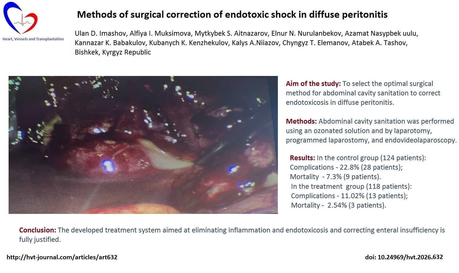
The aim of the study was to select the optimal surgical method for abdominal cavity sanitation to correct endotoxicosis in patients with diffuse peritonitis.
Methods
Study design: retrospective observational study.
The results of treatment of 242 patients with diffuse peritonitis aged 16 to 83 years who were admitted to the surgical department No. 1 of the City Clinical Hospital No. 1 in Bishkek for inpatient treatment between 2003 and 2025 were analyzed. Inclusion criteria: Patients with acute diseases of the abdominal organs complicated by diffuse secondary and tertiary peritonitis. Exclusion criteria: Patients who underwent repeat surgery for malignant neoplasms of the abdominal organs complicated by peritonitis, acute destructive forms of pancreatitis.
Using approaches to the treatment of diffuse peritonitis and analyzing the results, two groups of patients were identified: control (124 patients) – who underwent fractional dialysis of the abdominal cavity with antiseptic solutions, either with the addition of antibiotics and traditional drainage, intramuscular and intravenous administration of antibiotics; and the main group (118 patients) - who underwent abdominal cavity sanitation with an ozonated solution and methods of laparotomy, programmed laparostomy, and endovideolaparoscopy, regional lymphostimulation, administration of ozonated solutions into the abdominal cavity, and its drainage using methods developed by us.
All patients provided written informed consent for all procedures and surgery. The study complied with the requirements of the Declaration of Helsinki 2024. The study was approved by an independent bioethics committee at the Kyrgyz State Medical Institute for Retraining and Advanced Training named after S.B. Daniyarov (protocol No. 2 dated March 29, 2024).
In evaluating the treatment results, we used baseline characteristics, clinical signs of the disease course, the nature of complications, and the dynamics of laboratory tests.
Baseline and clinical course variables
We collected demographic and clinical data: age, sex, causes of peritonitis, comorbidities, laboratory tests (total blood count, electrolytes, bilirubin, alanine aminotransferase (ALT), aspartate amintotransferase (AST), urea, creatinine) and body temperature, peritoneal symptoms, passage of gases , stool, blood pressure, pulse rate, and duration of hospitalization.
Surgery
After brief preoperative preparation aimed at correcting volume disorders, protein and electrolyte imbalances, and restoring the impaired functions of major organs and systems, the patients underwent emergency surgery under endotracheal anesthesia.
In the control group, surgery was performed via a median laparotomy approach, which provided adequate revision and sanitation of the abdominal organs. However, it was not always possible to achieve the desired correction of impaired functions: there were cases of persistent peritonitis despite the causes being eliminated, encapsulated abscesses developed, and early adhesive intestinal obstructions occurred, resulting in fatal outcomes.
In this regard, in the main group, methods of programmed laparostomy, relaparotomy, and endovideolaparoscopy were used. The scope of the intervention included elimination of the source of peritonitis, sanitation of the abdominal cavity, and its drainage. To correct enteral insufficiency, small intestine intubation was performed using a double-lumen tube, which allowed the evacuation of toxic contents and enteral nutrition. The main parameters (cause, duration, and prevalence of the inflammatory process) in the groups were equivalent, but the course of the disease differed.
Most patients with perforated gastroduodenal ulcers underwent suturing due to late admission (Fig. 1).
In cases of peritonitis of any etiology, it is necessary to perform tests that would allow assessing the degree of inflammation, the severity of endotoxicosis, and the bacterial contamination of the abdominal cavity. These same tests, when performed dynamically, allow assessing the effectiveness of the treatment. With this in mind, in addition to general clinical tests (complete blood count, urinalysis, blood glucose, bilirubin, thymol test, ALT, AST, residual nitrogen, urea, creatinine, ECG), we also used special examination methods.
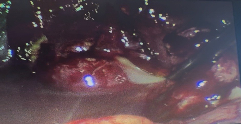
Figure 1. Laparoscopic abdominal cavity sanitation
Ultrasound examination and endovideolaparoscopy
For the purpose of diagnosis, and in some cases treatment in combination with objective and laboratory test data, ultrasound and endovideolaparoscopy were used, which were performed using Toshiba SaL and Olympus devices (Japan), respectively, when patients were admitted to the hospital, and these techniques were especially used in the postoperative period to diagnose intra-abdominal complications (abscesses, intestinal obstruction), and were also important in determining postoperative peritonitis.
During ultrasound, attention was paid to the diameter of the small intestine, the thickness of the wall, and the presence of fluid in its lumen. In cases of paralytic intestinal obstruction: an increase in the diameter of the intestine to 5-6 cm was noted, as well as thickening of its wall, intestinal villi, and converging valves in the form of a “keyboard” or “ladder,” and the presence of a large amount of fluid in the lumen with pendulum-like movement of intestinal contents synchronous with respiratory movements.
Assessment of the severity of endotoxicosis
To determine the severity of endotoxicosis, the following were used:
LII – leukocyte intoxication index determined according to Kalif-Kalif (15) using the formula:
LII = ((4 mts + 3yu + 2p + s) x (pl.c + 1)) / ((mon. + lymph.) x (e + 1))
where mts – myelocytes, yu – young cells, p – rod-shaped cells, s – segmented cells, pl.kl. – plasma cells, mon. – monocytes, lymph. – lymphocytes, e – eosinophils.
MSM – medium molecular weight molecules were determined using the method proposed by Gabrielian et al. (16) for rapid diagnosis.
PT – paramecium test for determining the toxicity of plasma and abdominal cavity exudate. The essence of the method is to determine the time (in minutes) of paramecium death.
Bacteriological evaluation and sanitation of abdominal cavity
When performing bacteriological examination of abdominal cavity exudate, we followed Order No. 535 of April 22, 1985, Moscow, and used the methods recommended by Menshikov (17). For bacteriological examination at the time of surgery, 2.0 ml of exudate was collected with a sterile syringe and transferred to a sterile test tube. During the period when drains were in place, exudate was collected by aspiration. Secretions from the wound were collected with a sterile cotton swab and placed in a sterile test tube. If anaerobic flora was suspected, the exudate or swab was placed in a test tube with thioglycolic semi-solid agar. The test material was cultured on 5% blood agar, sugar broth, egg yolk-salt agar, and thioglycolic medium.
The degree of contamination was assessed using the sector culture method: doubtful degree – 104 CFU/ml of microbial bodies in 1.0 ml, 105 CFU/ml – low degree, from 105 to 107 CFU/ml – medium degree, and above 107 CFU/ml – high degree of contamination.
To eliminate the inflammatory process in diffuse purulent peritonitis, we performed sanitation of the abdominal cavity with ozonated saline solution at an ozone concentration of 8-10 μg/ml. A significant aspect of our work was the determination of the microflora of the abdominal cavity exudate, which we took for examination during laparotomy or laparoscopy, and from drainage tubes in the postoperative period.
The abdominal cavity was washed with an ozonated solution during surgery, and in the postoperative period, it was introduced into the cavity through the upper safety drains, which prevented the formation of adhesions and the development of early adhesive intestinal obstruction. The dialysate drained by gravity through the lower safety drains located in the pelvic cavity.
The quality of abdominal cavity sanitation was assessed by bacterial culture of the exudate. If the bacterial culture was negative after treatment and on the third day, the result was considered good. If the culture was negative after treatment, but a monoculture with a low degree of contamination was detected on the third day, the result was considered satisfactory, and unsatisfactory if the culture on the first and third days revealed microflora with a moderate degree of contamination.
Follow-up and outcomes
The blood tests (total blood count, bilirubin, ALT, AST, urea, creatinine) and tests for endotoxicosis (LII, MSM, PT ) were performed before surgery, 1st day, 3rd day, 5-7 days and 10-12 days after surgery.
We also monitored days of normalization of body temperature, day of peritoneal symptoms disappearance, passage of gases, stool, normalization of blood pressure and pulse rate, and duration of hospitalization.
We recorded development of complications and mortality.
Statistical analysis
Statistical analysis was performed using Microsoft Excel and IBM SPSS Statistics 26.0. Data are presented and mean (SE) and number (%). Categorical variables were compared using Chi-square test and continuous variables using unpaired t-test for independent samples. A p value less than 0.05 was accepted as significant.
Results
Patients characteristics
The age of patients ranged from 16 to 83 years, with more men (161) than women (81) admitted. In the study, 22.7% (55 patients) belonged to the 21-30 years age group; 23.14% (56 patients) belonged to the 31-40 age group; 18.18% (44 patients) were aged 41-50; 9.0% (22 patients) were aged 51-60; and 16.95% (41 patients) were aged 61 and older.
Perforated stomach and duodenal ulcers, acute destructive forms of appendicitis and cholecystitis, and intestinal obstruction were the main causes of secondary diffuse peritonitis during the first operation.
The causes of relaparostomies, relaparoscopies, and relaparotomies in tertiary peritonitis were bile leakage from an improperly sutured choledochostomy tube, abscesses that ruptured into the free abdominal cavity, failure of gastrointestinal and interintestinal anastomosis sutures and the appendix stump, as well as ongoing peritonitis.
The comorbidities were present in 135 of 242 patients and it is known that their timely correction affects the outcome of the disease (Table 1).
|
Table 1. Nosological forms of comorbidities in patients with peritonitis and their frequency |
||
|
Comorbidities |
Total |
|
|
Abs. number |
% |
|
|
Coronary heart disease |
19 |
7.85 |
|
Diabetes mellitus |
5 |
2.07 |
|
Hypertension |
28 |
11.57 |
|
Pneumosclerosis |
8 |
3.30 |
|
Chronic bronchitis |
9 |
3.71 |
|
General atherosclerosis |
26 |
10.74 |
|
Pyelonephritis |
3 |
1.23 |
|
Obesity |
14 |
5.78 |
|
Varicose veins of the lower extremities |
4 |
1.65 |
|
Combination of diseases |
19 |
7.85 |
|
Total |
135 |
55.78 |
The bacteriological studies were performed in 118 patients out of 242 who underwent surgery, and the following microflora was isolated: E. coli – 82 (69.50%), Streptococcus spp. – 17 (14.41%), Bacteroides – 13 (11.02%), B. Fragilis – 3 (2.54%), Fusobacterium – 3 (2.54%).
Monoculture was found in 66 (55.93%) patients, and a combination of microflora in 52 (44.07%). Moreover, during the initial culture, the contamination level in all examined patients was above critical (108 CFU/ml).
Efficacy of sanitation of abdominal cavity
Surgical methods of sanitation such as programmed laparostomy (5.93%), relaparotomy (25.42%), and programmed endovideolaparoscopy (9.32%) were used.
The main focus was on body temperature dynamics. It was found that when using programmed laparoscopy for sanitation, body temperature returned to normal after the second abdominal sanitation, while with relaparostomy and relaparotomy, hyperthermia remained at hectic levels for at least 5-6 days and later on, it was elevated, although not above 37.5°C.
The second important sign that caught our attention was pain syndrome and early activation of patients, which are interrelated. With relaparoscopy, pain was not very pronounced after the first procedure and in the subsequent ones, which allowed patients to become active within five to six hours after the operation. With relaparostomy and relaparotomy, pain syndrome persisted throughout the treatment, which led to immobility, revealing the essence of the next indicator.
Early restoration of bowel function was important in the outcome of the disease. With relaparoscopy, function was restored in 2-3 days, while with relaparostomy and relaparotomy it took at least 4-5 days. The length of hospital stay was also important in evaluating the methods: with relaparoscopy, it was up to 16-18 days, while with relaparostomy and relaparotomy, it exceeded 28-30 days.
We compared the main clinical manifestations in the postoperative period in both groups (Table 2) and found that with the treatment we developed, clinical indicators improved significantly faster (p<0.05).
|
Table 2. Clinical indicators of patients with peritonitis |
|||
|
Variables |
Control group (n=124) |
Main group (n=118) |
p |
|
Normalization of body temperature, days |
5.2 (1.11) |
3.1(0.21) |
0.0706 |
|
Disappearance of peritoneal symptoms, days |
4.7(0.71) |
2.9(0.17) |
0.0167 |
|
Passage of gases, days |
5.7(0.74) |
3.1(0.32) |
0.0017 |
|
Stool, days |
6.1(0.55) |
3.4(0.42) |
0.0001 |
|
Blood pressure stabilization, days |
3.7(1.12) |
2.1(0.08) |
0.1657 |
|
Pulse rate stabilization, days |
4.8(1.21) |
2.7(0.14) |
0.09 |
|
Duration of hospitalization, days |
24.5(3.1) |
13.7(2.1) |
0.0047 |
|
Data are presented as mean (SE) t test for independent samples |
|||
Based on the analysis of the techniques used in the most severe patients admitted in the terminal stage of the disease, we developed criteria for determining the indications for repeat laparostomies, relaparotomies, reendovideolaparoscopies, and abdominal cavity sanitation in cases of diffuse peritonitis. These include peritoneal edema and lack of shine, a significant amount of purulent effusion with a putrid odor, the presence of tightly fixed fibrin, pronounced edema of the intestinal wall, and lack of peristalsis.
When relaparostomy, relaparotomy, and relaparoscopy were used, all patients had purulent effusion in the abdominal cavity with a putrid odor during the primary operation, the intestinal loops were covered with fixed gray fibrin deposits, the intestine was sharply distended throughout, its walls were edematous, and peristalsis was not detected. After removal of the focus of inflammation, the abdominal cavity was thoroughly treated two or three times with ozonated or twice-irradiated solutions with an exposure time of 5-10 minutes. We prefer to fill the abdominal cavity 2-3 times with solutions with an exposure time of 5-10 minutes rather than multiple rinsing, which is more traumatic. The operation was completed by fixing a microirrigator in the round ligament of the liver for the introduction of a lymphotropic mixture, followed by removal through an additional incision in the left hypochondrium and tight fixation to the skin so that it would not fall out during repeated sanitation. The abdominal cavity was then drained using a technique developed by us, after which in some patients who underwent programmed relaparostomy, rubber strips were applied to the intestine, and sutures were applied only to the skin; the subcutaneous tissue was drained, which ensured the outflow of wound exudate.
Relaparostomy was performed the day after the operation, then again 24 hours later.
The sutures on the skin were removed, the edges of the wound were spread apart, and the abdominal cavity was irrigated profusely with an ozonated solution through the drains.
It should be noted that after 24 hours, the drains became clogged with fibrin, so during repeated sanitation, they were necessarily washed, then the abdominal cavity was very carefully drained, and solutions were administered twice a day through the remaining drains. We repeated this procedure twice in four patients, three times in four others, and four times in three others.
The number of relaparostomies, relaparotomies, and reendoscopic laparoscopies was determined by the condition of the abdominal cavity, the nature of the effusion, the paramecium test of the exudate, and bacterial culture of the discharge from the abdominal cavity. The operations were stopped as soon as the peritoneal edema decreased, gloss and visible peristalsis appeared, there was a small amount of effusion in the abdominal cavity, no new areas of plaque formed, and the old ones were easily separated and removed. Then, narrow-spectrum infrared irradiation of the abdominal cavity was used.
It should be noted that, starting from the third day, the general condition of the patients gradually improved, the intestines were cleared of deposits, the effusion became serous-purulent and then serous, and the temperature returned to normal. A positive fact is that none of the patients experienced progression of peritonitis or other complications, despite the severity of their condition.
A significant improvement in endotoxicosis, liver, and kidney test results was observed with programmed endovideolaparoscopic abdominal sanitation. The general condition of the patient improves much faster and requires less attention in care. After a few applications of the relaparostomy method, we refrained from its further use and gave preference to endovideolaparoscopic interventions.
The decision to discontinue or continue repeated abdominal sanitation was difficult. During follow-up ultrasound examinations, we assessed the amount of effusion, the nature of the plaque on the internal organs, and the condition of the intestines, paying attention to endotoxicosis indicators, which remain high during treatment and do not reach normal levels by the 10th-12th day of the study (Table 3).
However, there were significant differences in endotoxicosis indices between groups. LII had statistically significant lower values in reendovideolaparoscopy group as compared to relaparoscopy group 1 week and 2 weeks after surgery (p<0.001 for both), as well as MSM (p<0.01, p<0.05 and p< 0.01, for 3rd day, 5-7th days and 10-12th days, respectively). While PT levels were higher at the same dates for reendovideolaparoscopy group as compared to relaparoscopy groups, approaching normal level on 10-12th day (p<0.01, p<0.001 and p<0.01, respectively).
|
Table 3. Dynamics of LII, MSM, and PT indicators in patients undergoing relaparoscopy and reendovideolaparoscopy |
|||||||
|
Variables |
Groups |
Examination period |
Normal values |
||||
|
Before surgery
|
1st Day
|
3rd Day
|
5-7th Day
|
10-12th Day
|
|||
|
LII |
1 |
5.12 (0.32) |
5.43 (0.27) |
4.28 (0.13) |
3.12 (0.61) |
2.13 (0.27) |
0.9(0.01)
|
|
2 |
5.16 (0.32 |
5.9 (0.99) |
3.05 (0.02) |
2.1 (0.03) |
1.2 (0.03) |
||
|
p |
|
>0.05 |
<0.05 |
<0.05 |
<0.01 |
<0.01 |
|
|
MSM, conditional units |
1 |
0.774 (0.012) |
0.702 (0.014) |
0.792(0.013) |
0.527(0.011) |
0.428 (0.012) |
0.239(0.01) |
|
2 |
0.76 (0.07)
|
0.70 (0.015)
|
0.61(0.003)
|
0.44 (0.02)
|
0.29 (0.009)
|
||
|
p |
|
>0.05 |
>0.05 |
<0.01 |
<0.05 |
<0.01 |
|
|
PT, min |
1 |
14.2 (0.13) |
12.7 (0.18) |
14.8 (0.11) |
18.6 (0.13) |
19.7 (0.12) |
26.0(0.21) |
|
2 |
13.7 (0.11) |
11.9 (0.06) |
16.1 (0.06) |
22.8 (0.03) |
24.8 (0.02) |
||
|
p |
|
<0.05 |
>0.05 |
<0.01 |
<0.001 |
<0.01 |
|
|
Data are presented as mean (SE), p – differences between relaparoscopy and reendovideolaparoscopy groups 1 - relaparoscopy and 2 - reendovideolaparoscopy t-test for independent samples LII –leukocyte intoxication index, MSM - medium molecular weight molecules, PT – paramecium test |
|||||||
Similar dynamics were observed in biochemical tests (Table 4). Positive dynamics were observed in bilirubin levels, with a decrease in bilirubin noted starting on the third day after relaparostomy, but even on the 12th day, it still differed from the normal value. However, in reendovideolaparoscopy group bilirubin reduced significantly approaching normal leel on 10-12th day and it levels were significantly lower on 5-7th days and 10-12th days as compared to relaparoscopy group (p<0.001 and p<0.05, respectively).
As for AST and ALT levels, they decreased significantly on day 3, remained the same as on day 3 on days 5-7, and then decreased. They were significantly lower in reendovideolaparoscopy group as com[pared relaparoscopy group on 5-7th day and 10-12th day (p<0.05 and p<0.01 for ALT, p<0.01 and p<0.001 for AST).
Urea and creatinine began to decrease only on days 5-7 and had not yet reached normal levels by day 12, but their level was significantly lower in reendovideolaparoscopy group as compared to relaparoscopy group (p<0.05 for 3rd, 507th and 10-12th days for both urea and creatinine).
Biochemical studies showed that in cases of diffuse peritonitis of any origin, liver and kidney functions are impaired, and during relaparostomy, relaparotomy, and relaparoscopy, the functional state of these organs improves to varying degrees, confirming the need for their use in the treatment of diffuse peritonitis.
|
Table 4. Dynamics of biochemical parameters in patients undergoing relaparostomy and reendovideolaparoscopy |
||||||
|
Variables |
Groups |
Examination period |
Normal values
|
|||
|
Before operation
|
3rd day
|
5-7th days
|
10-12th days
|
|||
|
Bilirubin, μmol/L |
1 |
34.5(1.12) |
27.4(0.31) |
24.2(0.14) |
20.3(0.11) |
11.2(1.92) |
|
2 |
32.3(0.97) |
24.3(0.27) |
19.4(0.18) |
13.4(0.41) |
||
|
p |
|
>0.05 |
>0.05 |
<0.001 |
<0.05 |
|
|
ALT, μkat/L |
1 |
0.14(0.009) |
0.17(0.007) |
0.14(0.008) |
0.11(0.007) |
0.08(0.01) |
|
2 |
0.16(0.089) |
0.15(0.067) |
0.10(0.062) |
0.09(0.001) |
||
|
p |
|
>0.05 |
>0.05 |
<0.05 |
<0.01 |
|
|
AST, μkat/L |
1 |
0.13(0.008) |
0.16(0.006) |
0.15(0.005) |
0.12(0.006) |
0.07(0.01) |
|
2 |
0.14(0.007) |
0.13(0.005) |
0.11(0.004) |
0.08(0.005) |
||
|
p |
|
>0.05 |
>0.05 |
<0.01 |
<0.001 |
|
|
Urea, mmol/L |
1 |
9.8(0.92) |
8.7(0.64) |
7.4(0.31) |
6.21(0.07) |
5.3(0.08) |
|
2 |
8.7(0.47) |
7.1(0.21) |
6.1(0.21) |
5.1(0.4) |
||
|
p |
|
>0.05 |
<0.05 |
<0.01 |
<0.05 |
|
|
Creatinine, μmol/L |
1 |
174.7(2.14) |
189.5(1.17) |
162.4(2.12) |
120.4(1.13) |
68.7(1.14) |
|
2 |
169.5(2.09) |
158.2(1.48) |
141.7(1.42) |
110.5(0.71) |
||
|
p |
|
>0.05 |
<0.005 |
<0.01 |
<0.05 |
|
|
Data are presented as mean (SE), p – differences between relaparoscopy and reendovideolaparoscopy groups 1 - relaparoscopy and 2 - reendovideolaparoscopy t-test for independent samples ALT – alanine aminotransferase, AST – aspartate aminotransferase |
||||||
Complications and mortality
Analysis of complications and mortality in both groups showed that the nature of complications was identical, but in the main group, complications were easily remedied. Thus, complications in the control group occurred in 28 patients (22.8%), of whom 9 (7.3%) died. As a result of prolonged intestinal paresis, 5 patients developed intestinal evisceration, which required repeat surgery, 2 developed gastroenterostomy failure, 6 more developed peritonitis, forming an interloop abscess, 6 developed respiratory failure due to lower lobe pneumonia, and 9 patients developed early adhesive intestinal obstruction, requiring repeat surgery.
In the main group, complications were noted in 13 (11.02%) patients, and 3 (2.54%) patients who were admitted to the hospital in already serious condition and died.
Comparison of complications rate demonstrated statistically significantly (p=0.025) lower complications rate in main group as compared to control one (11.02% vs. 22.8%), however mortality showed a trend to lower values in main group (2.54% vs. 7.3%, p=0.091). These results confirm the appropriateness of the treatment we used for patients with peritonitis.
Discussion
Our study demonstrated that developed treatment of endotoxicosis in peritonitis facilitated normalization of body temperature, disappearance of symptoms and restoration of bowel activity, markedly reduced hospital stay. It decreased indexes of endotoxicosis, improved biochemical liver and renal parameters, and significantly reduced complications rate and mortality in patients with peritonitis.
Peritonitis remains a common and dangerous complication of acute diseases of the abdominal organs. Advanced age, comorbidities, disruption of the digestive tract seal, and delayed surgery are the main factors contributing to high mortality (5). The key to treating peritonitis is to eliminate the source of peritonitis or effectively isolate it from the free abdominal cavity, adequate sanitation, and drainage of the abdominal cavity (1).
Analysis of the diseases showed that the most common causes of peritonitis were perforated stomach and duodenal ulcers (58.54%), destructive forms of appendicitis (29.27%), and acute intestinal obstruction (12.19%). It should be noted that in 90% of cases, patients with fatal outcomes in widespread peritonitis were admitted to the surgical hospital more than 24 hours after the onset of the disease, which was due to the progression of peritoneal inflammation and multiple organ failure. The age of patients is also a critical factor influencing the course of abdominal sepsis.
The use of minimally invasive technologies in the treatment of diffuse peritonitis has reduced the number of postoperative complications and the level of postoperative mortality (18-20). Our results confirmed reduction of complications and mortality.
Thus, diagnostic endovideolaparoscopy made it possible to establish a diagnosis, determine treatment tactics, and perform abdominal cavity sanitation in 24.39% of patients, with 4.88% of them being eligible for laparoscopic suturing of perforated gastroduodenal ulcers. The transition to conversion when it was necessary to perform surgery through laparotomy was also considered positive (9, 21).
Study limitations
This study has a limitation that should be taken into account when interpreting the results: its retrospective design. The analysis was performed using existing medical records, which limits the ability to take all factors into account.
Conclusions
Endovideolaparoscopy was used in the diagnosis of diffuse peritonitis, and programmed laparostomy, programmed laparoscopy, and relaparotomy were used in treatment. Treatment of the abdominal cavity with ozonated solutions and their introduction into the abdominal cavity in the postoperative period contribute to faster elimination of bacterial contamination. The use of the treatment complex we have developed ensures the elimination of peritoneal inflammation and restoration of bowel function. The assessment was made using clinical, laboratory, and instrumental research results. The advantages of endovideolaparoscopy have been convincingly demonstrated, on the basis of which this method can be recommended for the treatment of diffuse peritonitis.
Ethics: All patients provided written informed consent for all procedures and surgery. The study complied with the requirements of the Declaration of Helsinki 2024. The study was approved by an independent Bioethics Committee at the Kyrgyz State Medical Institute for Retraining and Advanced Training named after S.B. Daniyarov (protocol No. 2 dated March 29, 2024).
Peer-review: External and internal
Conflict of interest: None to declare
Authorship: Study concept and design – U.D.I., A.I.M, M.S.AM; Data collection and processing – U.D.I, A.I.M; Statistical analysis – A.I.M., E.N.N., A.N.u., M.S.A., K.K.B., K.K.K., K.A.N., Ch.T.E., A.A.T.; Text writing – U.D.I., A.I.M., E.N.N., A.N.u., M.S.A., K.K.B., K.K.K., K.A.N., Ch.T.E., A.A.T.; Editing – U.D.I., A.I.M., M.S.A. Thus all authors equally contributed to preparation of manuscript and fulfilled all authorship criteria
Acknowledgements and funding: None to declare
Statement on A.I.-assisted technologies use: The authors did not use AI-supported technologies in the preparation of this manuscript.
Data and material availability: Contact authors. Any share should be in frame of fair use with acknowledgement of source and collaboration.
References
| 1. Matveev IA, Dmitriev AV, Abrahamyan KV. Postoperative peritonitis. Epidemiology, diagnosis, surgical treatment, and prognosis. Surgery (Journal named after N.I. Pirogov) 2025; 6: 104-11. doi: 10.17116/hirurgia2025061104 https://doi.org/10.17116/hirurgia2025061104 PMid:40455026 |
||||
| 2. Gorbulich AV, Chernikova AA, Bilyalova DE, Savchenko DA. Peritonitis. Integr Med Educ 2024; 4: 27-9. | ||||
| 3. Asan TB, Baltabaeva UB, Begaly AK, Kaiyp NM, Rahman NN, Sarybai AA, et al. Main causes of death in widespread peritonitis. Bulletin of KazNMU 2020; 4: 349-54. | ||||
| 4. Mamakeev KM, Alybaev EU, Sadabaev TYu, Mamakeev ZhB. Optimization of surgical treatment tactics for perforated pyloroduodenal ulcers in elderly and senile patients. Bull Sci Pract 2022; 8: 242-50. doi: 10.33619/2414-2948/76/26 https://doi.org/10.33619/2414-2948/76/26 |
||||
| 5. Sartelli M, Catena F, Ansaloni L, Poiasina E, Coccolini F, Moore E, et al. Complicated intra-abdominal infections in a worldwide context: an observational prospective study (CIAOW Study). World J Emerg Surg 2013; 8: 22-6. https://doi.org/10.1186/1749-7922-8-1 PMid:23286785 PMCid:PMC3538624 |
||||
| 6. Kurbanbaeva G, Ismailov F. Peritonitis in elderly and senile patients. Sci Works Gift Youth Med 21st Cent 2023; 1: 106. | ||||
| 7. Chyngysheva ZhA, Niyazov BS, Naziraliev R, Adylbaeva VA, Dinlosan OR, Abdullaev ZhS. A modern view on the diagnosis and treatment of acute intestinal obstruction in geriatric patients. Bull Sci Pract 2022; 8: 261-92. doi: 10.33619/2414-2948/80/2 https://doi.org/10.33619/2414-2948/80/24 |
||||
| 8. Balayan AZ. Clinical features of acute cholecystitis in the elderly. Exp Clin Gastroenterol 2016; 6: 60-4. | ||||
| 9. Ermolov AS, Samsonov VT, Yartsev PA, Gulyayev AA. Video laparoscopic diagnosis and surgical tactics for acute diseases of the abdominal organs. Medical Alphabet 2021; 4: 17-24. doi: 10.33667/2078-5631-2021-4-17-24 https://doi.org/10.33667/2078-5631-2021-4-17-24 |
||||
| 10. Sopuev AA, Kudaiberdiev ZK, Umurzakov OA, Mamytov KN, Mambetov AK. Factors of perforation risk in acute appendicitis in elderly and senile patients. Med Sci 2021; 5: 5-11. | ||||
| 11. Sattarov ShT, Ruzibaev SA, Khursanov EE. Optimization of the path of correction of endotoxicosis in acute peritonitis. Res Focus 2022; 2: 144-50. | ||||
| 12. Gasanov MD. Formation of algorithms for determining the severity of endotoxicosis in peritonitis. Surgery (Journal named after N.I. Pirogov) 2015; 1: 54-7. https://doi.org/10.17116/hirurgia2015154-57 PMid:25909553 |
||||
| 13. Saraev AR, Nazarov ShK. Pathogenesis and classification of widespread peritonitis. Surgery (Journal named after N.I. Pirogov) 2019; 12: 106-10. doi: 10.17116/hirurgia2019121106 https://doi.org/10.17116/hirurgia2019121106 PMid:31825350 |
||||
| 14. Grachev V, Marinkin I, Batyrev V. Abdominal pain. J Sci (Lyon) 2021; 24: 19-30. | ||||
| 15. Kalif-Kalif YY. On the leucocyte index of intoxication and its prognostic value. Med Bus 1941; 1: 31-5. | ||||
| 16. Gabrelian NI, Dmitriev AA, Kulakov GP, Mekikian AM, Scherbanova OI. Diagnostic value of determination of middle molecules in blood plasma in nephorlogical diseases. Klin Med 1981; 59: 38-42. | ||||
| 17. Menshikov VV. Laboratory Methods of Examination in the Clinic. Moscow, 1987. 565p. | ||||
| 18. Glushkov NI, Kabanov MYu, Gorshenin TL, Semencov KV, Lobanov MYu, Dulaeva SK. Improving the diagnosis and treatment tactics for elderly and senile patients with acute obstructive colonic obstruction of tumor origin. Adv Gerontol 2020; 33: 908-15. doi: 10.34922/AE.2020.33.5.011 https://doi.org/10.34922/AE.2020.33.5.011 |
||||
| 19. Abdulzhalilov MK, Saiddiibirov ShM. Evaluation of the effectiveness of modern methods of surgical treatment of patients with perforated gastric and duodenal ulcers. Bull Dagestan State Med Acad 2022; 43; 70-7. | ||||
| 20. Ziyev ShKh, Daminova F Kh, Nozimov KYu, Obidzhoni NM. The current state of problems in the surgical treatment of acute cholecystitis in elderly and senile patients. Bull Pedagog Univ Nat Sci 2021; 1-2: 295-300. | ||||
| 21. Rakhmatullaev RR, Kurbonov DD, Juraeva NKh, Rahmatullaev AR, Rahmatullaeva FR, Salihov RD. Surgical tactics for destructive cholecystitis in elderly and senile patients. Sci Innov 2024; 4: 80-6. | ||||
Copyright

This work is licensed under a Creative Commons Attribution-NonCommercial 4.0 International License.
AUTHOR'S CORNER