Comparison of AHA/ACC and ESC guidelines for the management of patients with stable coronary heart disease: same goal, unique perspectives
EDITORIALS
Comparison of AHA/ACC and ESC guidelines for the management of patients with stable coronary heart disease: same goal, unique perspectives
Article Summary
- DOI: 10.24969/hvt.2024.523
- CARDIOVASCULAR DISEASES
- Published: 26/10/2024
- Received: 21/10/2024
- Accepted: 21/10/2024
- Views: 14303
- Downloads: 2560
- Keywords: editorial
Address for Correspondence: Andrea Lorenzo, Institute of Cardiology, Rio de Janeiro, Brazil
Email: andlorenzo@hotmail.com
ORCID: 0000-0001-8522-6612
Andrea Lorenzo, Institute of Cardiology, Rio de Janeiro, Brazil
Abstract
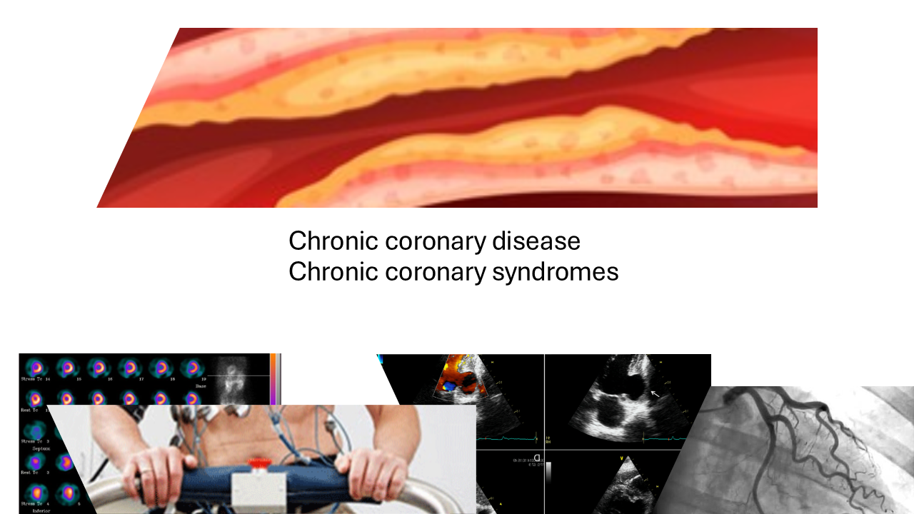
Currently, there are two updated guidelines (The 2023 AHA/ACC/ACCP/ASPC/NLA/PCNA and the 2024 European Society of Cardiology Guidelines) (1,2) dedicated to stable coronary artery disease (CAD), with different definitions. While the first defines chronic coronary disease, the latter employs a different terminology, chronic coronary syndromes, based on expanded pathophysiological concepts, including microcirculatory disease. Nonetheless, both are devoted to offering insights into the best patient management strategies available. Regarding diagnostic testing, both guidelines stress the importance of clinical and risk factor evaluation for the assessment of the likelihood of CAD, which will influence the choice of noninvasive tests- either anatomic or functional, along with specific patient characteristics that may affect the performance of each type of noninvasive test, as well as local expertise and test availability.
Graphical abstract

Currently, there are two updated guidelines (The 2023 AHA/ACC/ACCP/ASPC/NLA/PCNA and the 2024 European Society of Cardiology Guidelines) (1, 2) dedicated to stable coronary artery disease (CAD)- with a wide range of recommendations, from the best available assessment strategies to treatments. Both provide evidence-based information, and the choice between guidelines may rely on regional practices and preferences.
To start, one should notice different definitions. The 2023 AHA/ACC guidelines define chronic coronary disease (CCD) as “patients discharged after an acute coronary syndrome or after coronary revascularization procedure; patients with left ventricular systolic dysfunction and known or suspected CAD or those with established cardiomyopathy deemed to be of ischemic origin; patients with stable angina symptoms or ischemic equivalents medically managed with or without positive results of an imaging test; patients with angina symptoms and evidence of coronary vasospasm or microvascular angina; patients diagnosed with CCD based solely on the results of a screening study and whose treating clinician concludes that the patient has CAD” (1). On the other hand, the ESC defines chronic coronary syndromes (CCS), describing the clinical presentations of CAD during stable periods, defining CAD as the pathological process characterized by atherosclerotic plaque accumulation in the epicardial arteries, whether obstructive or non-obstructive. Based on expanded pathophysiological concepts, a more comprehensive definition of CCS was introduced: “a range of clinical presentations or syndromes that arise due to structural and/or functional alterations related to chronic diseases of the coronary arteries and/or microcirculation. These alterations can lead to transient, reversible, myocardial demand vs. blood supply mismatch resulting in hypoperfusion (ischemia), usually (but not always) provoked by exertion, emotion or other stress, and may manifest as angina, other chest discomfort, or dyspnea, or be asymptomatic.
Although stable for long periods, chronic coronary diseases are frequently progressive and may destabilize at any moment with the development of an acute coronary syndrome” (2).
Regarding testing, according to the AHA guidelines, in patients with CCD it is recommended that risk stratification incorporate all available information, including noninvasive, invasive, or both cardiovascular diagnostic testing results or use validated risk scores to classify patients as low (<1%), intermediate (1%-3%), or high (>3%) yearly risk for cardiovascular death or nonfatal myocardial infraction (MI).
If possible, medical treatment should first be intensified and testing deferred. Imaging should be considered in those with new-onset or persistent stable chest pain. Assessing the severity of ischemia may be useful to guide clinical decision-making regarding the use of invasive coronary angiography (ICA). In patients with CCD and frequent angina or severe stress-induced ischemia, referral to ICA or coronary computed tomography angiography (CCTA) is an option.
In patients with CCD and a change in symptoms or functional capacity that persists despite optimized medical treatment, stress positron emission tomography (PET)/single photon emission computed tomography myocardial perfusion imaging (SPECT MPI), cardiovascular magnetic resonance (CMR) imaging, or stress echocardiography are recommended to detect the presence and extent of myocardial ischemia, estimate risk of major adverse cardiovascular events, and guide therapeutic decision-making. The guidelines provide detailed information about the different modalities of diagnostic tests, their indications and contraindications.
In patients with CCD with newly reduced left ventricular systolic function, clinical heart failure, or both, ICA is recommended to assess coronary anatomy and guide potential revascularization. ICA for risk stratification is not routinely recommended in patients without left ventricular systolic dysfunction, heart failure, stable chest pain refractory to optimized medical treatment, and/or noninvasive testing suggestive of significant (>50%) left main disease.
Regarding testing for the assessment of CCS, the 2024 ESC guideline recommends starting with clinical evaluation, a 12-lead resting electrocardiogram, basic blood tests, chest X-ray imaging and pulmonary function testing in selected individuals, followed by echocardiography at rest to rule out left ventricular dysfunction or valvular heart disease, and exercise stress testing. The estimation of the clinical likelihood of obstructive CAD guides further noninvasive or invasive testing, employed to establish the diagnosis of CCS and to determine the risk of adverse events.
In the case of very low clinical likelihood of obstructive CAD (<5%), deferring further testing may be considered, unless symptoms persist and other noncardiac causes have been excluded. That applies also to patients with severe comorbidities, frailty of reduced life expectancy, who may proceed to medical treatment. In individuals with low (>5% - 15%) pre-test likelihood of obstructive CAD, coronary artery calcium score (CACS) should be considered to reclassify subjects and to identify more individuals with very low (≤5%) CACS-weighted clinical likelihood. In patients with a low (>5%–15%) likelihood of obstructive CAD, the benefit of diagnostic testing is uncertain but may be performed if symptoms are limiting and require clarification. Patients with moderate (>15%–50%), high (>50%–85%), and very high (>85%) likelihood of obstructive CAD should undergo further diagnostic testing.
Further non-invasive testing is, most of the times, either anatomic or functional noninvasive imaging, the choice of which depends on the pretest likelihood of obstructive CAD, patient characteristics that influence the performance of each type of noninvasive test (ie, comorbidities, obesity, left bundle branch block or other conditions which may affect image quality in some imaging modalities), as well as local expertise and availability. In individuals with low to moderate (>5–50%) clinical likelihood, CCTA is currently preferred to rule out obstructive CAD and detect nonobstructive CAD. On the other hand, in individuals with moderate to high (>15–85%) clinical likelihood, functional imaging such as myocardial perfusion scintigraphy, stress echocardiography or stress cardiac magnetic resonance perfusion, which assess the presence, severity and extent of myocardial ischemia, are useful for symptom correlation and guiding decisions on coronary revascularization. Positron emission tomography is ideal for absolute myocardial blood flow measurements, while cardiac magnetic resonance may be also used. The combined use of anatomic and functional may help decisions in patients with abnormal CCTA or abnormal functional testing, to improve patient selection for ICA.
ICA is recommended to diagnose obstructive CAD in individuals with a very high clinical likelihood (>85%), severe symptoms refractory to guideline-directed medical therapy, angina at a low level of exercise, suspicion of high-risk obstructive CAD, or severe myocardial ischemia.
Additionally, ICA with the availability of invasive functional assessments is recommended to confirm or exclude the diagnosis of obstructive CAD or angina/ischemia with no obstructive CAD (ANOCA/INOCA) in individuals with an uncertain diagnosis on non-invasive testing.
In the specific subset of patient with suspected ANOCA/INOCA (individuals with symptoms suggestive of myocardial ischemia and coronary arteries that are either normal or with non-obstructive lesions on CCTA or ICA), coronary blood flow quantification may be useful. Microvascular function assessment may be achieved by PET myocardial perfusion imaging, as well as dynamic SPECT MPI with coronary flow reserve assessment in new gamma-cameras or cardiac magnetic resonance.
In conclusion, both guidelines are valuable aids for the assessment and management of patients with stable CAD. Following either one or the other will ultimately set the path for the improvement of the outcomes of these patients.
Peer-review: Internal
Conflict of interest: None to declare
Authorship: A.L
Acknowledgement and Funding: None to declare
Statement on A.I.-assisted technologies use: Author declared they did not use A.I.- assisted technologies in preparation of manuscript
Availability of data and material: Do not apply
References
- 1.Virani SS, Newby LK, Arnold SV, Bittner V, Brewer LC, Demeter SH, et al.; Peer Review Committee Members. 2023 AHA/ACC/ACCP/ASPC/NLA/PCNA Guideline for the Management of Patients with Chronic Coronary Disease: A Report of the American Heart Association/American College of Cardiology Joint Committee on Clinical Practice Guidelines. Circulation 2023; 148: e9-e119. doi: 10.1161/CIR.0000000000001168
- 2. Vrints C, Andreotti F, Koskinas KC, Rossello X, Adamo M, Ainslie J, Banning AP, Budaj A, Buechel RR, Chiariello GA, Chieffo A, Christodorescu RM, Deaton C, Doenst T, Jones HW, Kunadian et al.; ESC Scientific Document Group. 2024 ESC Guidelines for the management of chronic coronary syndromes. Eur Heart J 2024; 45: 3415-37. doi: 10.1093/eurheartj/ehae177
Copyright

This work is licensed under a Creative Commons Attribution-NonCommercial 4.0 International License.
AUTHOR'S CORNER

